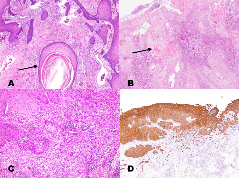
Fig 1.
a. A keratinizing (well-differentiated) squamous cell carcinoma showing large keratin pearls (arrows) and well-defined tumor cell outlines with slender cytoplasmic connections (intercellular bridges). Nuclear pleomorphism was mild, and the mitotic rate was low. b. Moderately differentiated OSCC (grade 2) presenting nests of basaloid tumor cells with peripheral palisading at the stromal interface. Intercellular bridges may have been present, but keratinization was focal (arrow). c. Poorly differentiated OSCC (grade 3) presenting confluent nests of tumor cells lacking keratinization or intercellular bridges. There is a slight background lymphocytic infiltrate, likely representing a host response. d. The identification of this grade 3 tumor as squamous carcinoma may require confirmatory immunostaining for cytokeratin, p63, or p40 (the same patient in Fig 1C with immunostaining for cytokeratin).

