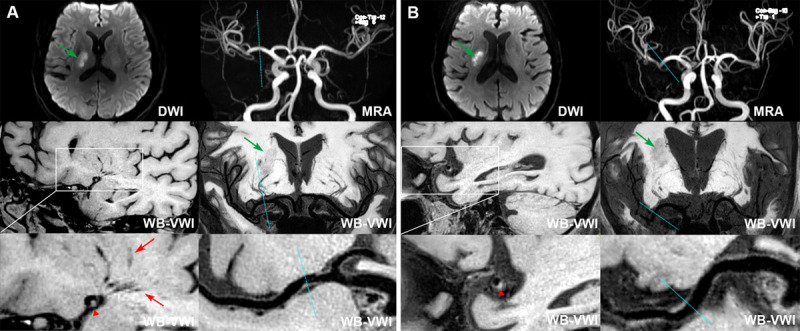
Figure 1.

Classification of proximal plaque and distal plaque on middle cerebral artery (MCA). Two representative cases of proximal plaque (located adjacent to the lenticulostriate artery [LSA] origin; (A) and distal plaque (located distal to the LSA origin; (B) on MCA. A, Diffusion-weighted imaging (DWI) and coronal minimum intensity projection (MinIP) showed a single subcortical infarction in the right LSA territory (green arrows); Magnetic resonance angiography (MRA) showed no stenosis on the relevant MCA; Coronal MinIP revealed shorter lengths of right LSAs compared with the left side and the curved multiplanar reconstruction (curved-MPR) showed a plaque adjacent to the corresponding LSA origins (blue dashed lines); corresponding cross-section view also demonstrated a traceable LSAs (red arrows) originating from the dorsally located plaque (arrowhead) of MCA. B, DWI and coronal MinIP showed a single subcortical infarction in the right LSA territory (green arrows); MRA showed no stenosis on the relevant MCA; Coronal MinIP revealed shorter lengths of right LSAs compared with the left side, but the curved-MPR showed a plaque distal to the corresponding LSA origin vessel segment of MCA (blue dashed lines); corresponding cross-section view demonstrated a dorsal plaque without traceable LSAs from the MCA wall.
