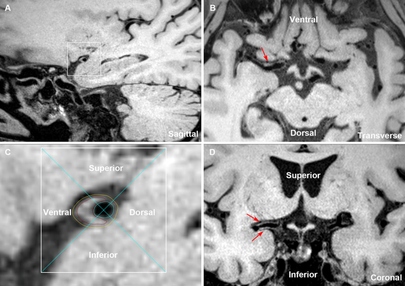
Figure 3.

The plaque distribution on middle cerebral artery (MCA) walls. A cross-section of plaque was divided into 4 quadrants (superior, inferior, ventral, and dorsal) using 2 perpendicular dashed lines that intersect at the lumen center, as shown by the enlarged image of plaque. The lumen area was outlined by blue lines, the outer wall area was outlined by yellow lines, and the estimated plaque was outlined by red lines (A and C). The reconstructed transverse and coronal views of MCA walls were used to further verify the involved quadrants of the plaque (B and D). The insets showed a typical plaque mainly involving the ventral, superior, and inferior wall (C, red lines; B and D, arrows).
