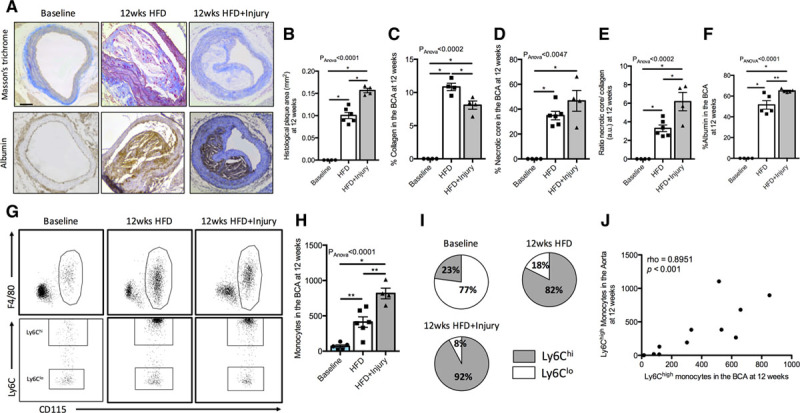
Figure 2.

Persistent arterial inflammation promotes formation of more inflamed, lipid-rich, and collagen-poor plaques in a remote arterial segment. A, Representative trichrome (upper row) and albumin immunohistochemistry (lower row) stainings of the different experimental groups. Quantification of the (B) plaque area, (C) collagen content, (D) necrotic core, (E) ratio of necrotic core to collagen content, and (F) albumin at 12 wk in all experimental groups (n=4/group). G, Representative flow cytometry images of the gating strategy employed to quantify monocytes in the brachiocephalic artery (BCA) and Ly6Chi and Ly6Clo monocyte subsets in all experimental groups at 12 wk. H, Flow cytometric quantification of monocytes in the BCA (n=6/group). I, Pie charts show the relative proportion of Ly6Chi and Ly6Clo monocytes in all the groups at the 12 wk time point (n=6/group). J, Correlation of Ly6Chi monocytes in the aorta with Ly6Chi monocytes in the BCA at the 12 wk time point. Data were represented as mean±SEM. For multiple-group comparisons, data were analyzed with a Kruskal-Wallis ANOVA with Dunn post hoc test. Correlation data were analyzed with a 2-tailed nonparametric Spearman test. HFD indicates high-fat diet. *P<0.05 and **P<0.01.
