
Figure 3.
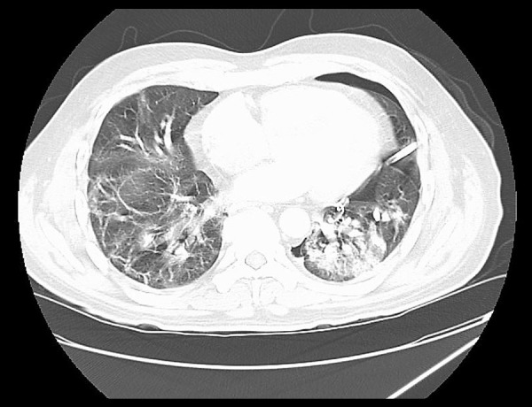
CT scan of the chest without contrast after pigtail insertion. Left pleural catheter is in position with approximately 27% volume left pneumothorax. Bilateral upper and lower lobe airspace infiltrates/consolidation are noted, left more pronounced than right.