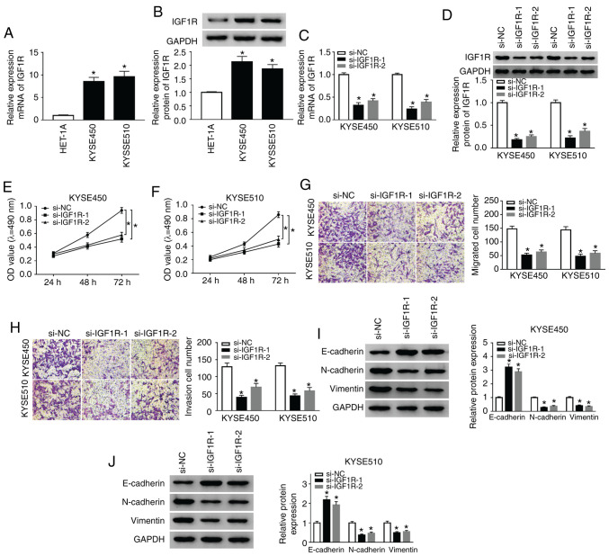
Figure 3.
Silencing of IGF1R suppresses the proliferation, migration, invasion and EMT of ESCC cells. (A) RT-qPCR was conducted to detect the expression of IGF1R in ESCC and HET-1A cells. (B) Western blot analysis of IGF1R protein expression in ESCC and HET-1A cells was implemented. (C-J) KYSE510 cells were transfected with si-NC, si-IGF1R-1, or si-IGF1R-2. (C and D) RT-qPCR or western blot analysis were used to assess the expression of IGF1R mRNA and protein in KYSE450 and KYSE510 cells. (E-H) MTT or Transwell assays was utilized for the determination of the proliferation, migration, and invasion of KYSE450 and KYSE510 cells. (I and J) The levels of E-cadherin, N-cadherin, and Vimentin in KYSE450 and KYSE510 cells were evaluated by western blot analysis. Data are presented as the means ± standard error of 3 experimental results. One-way ANOVA was applied to compare differences between groups. *P<0.05. BANCR, BRAF activated non-coding RNA; IGF1R, insulin-like growth factor 1 receptor; ESCC, esophageal squamous cell carcinoma.

