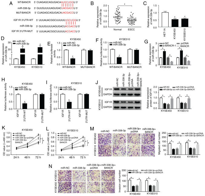
Figure 5.
BANCR modulates IGF1R expression via sponging miR-338-3p. (A) The potential binding sites between miR-338-3p and BANCR or IGF1R were predicted using starBase2.0. (B and C) The expression of miR-338-3p in ESCC tissues and adjacent normal tissues, as well as ESCC cells and HET-1A cells, was detected by RT-qPCR. (D) Following miR-NC or miR-338-3p transfection, the levels of miR-338-3p in KYSE450 and KYSE510 cells were detected by RT-qPCR. (E and F) Dual-luciferase reporter assay was conducted for the determination of the luciferase activity in KYSE450 and KYSE510 cells co-trans-fected luciferase reporters containing WT-BANCR or MUT-BANCR with miR-NC or miR-338-3p. (G) Effects of BANCR knockdown or overexpression on the expression of miR-338-3p were determined by RT-qPCR. (H and I) The luciferase activity of luciferase reporters containing IGF1R 3′UTR-WT or IGF1R 3′UTR-MUT in KYSE450 and KYSE510 cells transfected with miR-NC or miR-338-3p was detected using dual-luciferase reporter assay. (J-N) KYSE450 and KYSE510 cells were transfected with miR-NC, miR-338-3p, miR-338-3p+cDNA, or miR-338-3p+BANCR. (J) The levels of IGF1R protein in KYSE450 and KYSE510 cells were measured via using western blotting. (K-N) MTT or Transwell assays was carried out to analyze the proliferation, migration, and invasion of KYSE450 and KYSE510 cells. Data are presented as the means ± standard error of 3 experimental results. The Student's t-test or one-way ANOVA were applied to compare differences between groups where appropriate. *P<0.05. BANCR, BRAF activated non-coding RNA; IGF1R, insulin-like growth factor 1 receptor; ESCC, esophageal squamous cell carcinoma.

