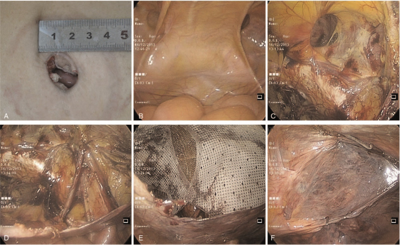
Figure 2.

Intraoperative laparoscopic photographs. (A) Umbilicus incision was made. (B) Exposure of the hernia defect. (C) Exposure of the pubic symphysis and Cooper's ligament. (D) Visualization of the spermatic cord and myopectineal orifice. (E) The mesh was placed to overlap the hernia opening. (F) Closure of the peritoneal defect with suture.
