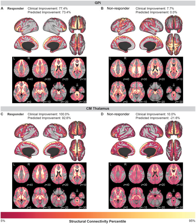
Figure 3.
Examples of tic improvement prediction for individual patients implanted in the GPi (top row) or the centromedial thalamus (bottom row). (A) A responder to GPi DBS with 77.4% improvement in tics showed high connectivity to limbic networks (anterior cingulate cortex, orbitofrontal cortex) and associative networks (medial and lateral prefrontal cortex) and low connectivity to sensorimotor networks (primary motor cortex, primary sensory cortex, and premotor areas). (B) In contrast, a non-responder to GPi DBS with only 7.7% improvement in tics showed high connectivity to sensorimotor networks and low connectivity to limbic and associative networks. (C) A responder to centromedial (CM) thalamus DBS with 100% improvement in tics showed high connectivity to sensorimotor networks (supplementary motor area, motor cortex, and sensory cortex) and lower connectivity to prefrontal and orbitofrontal areas. (D) In contrast, a non-responder to centromedial thalamus DBS with only 10.0% improvement in tics showed low connectivity to sensory and motor cortices and higher connectivity to prefrontal and orbitofrontal areas.

