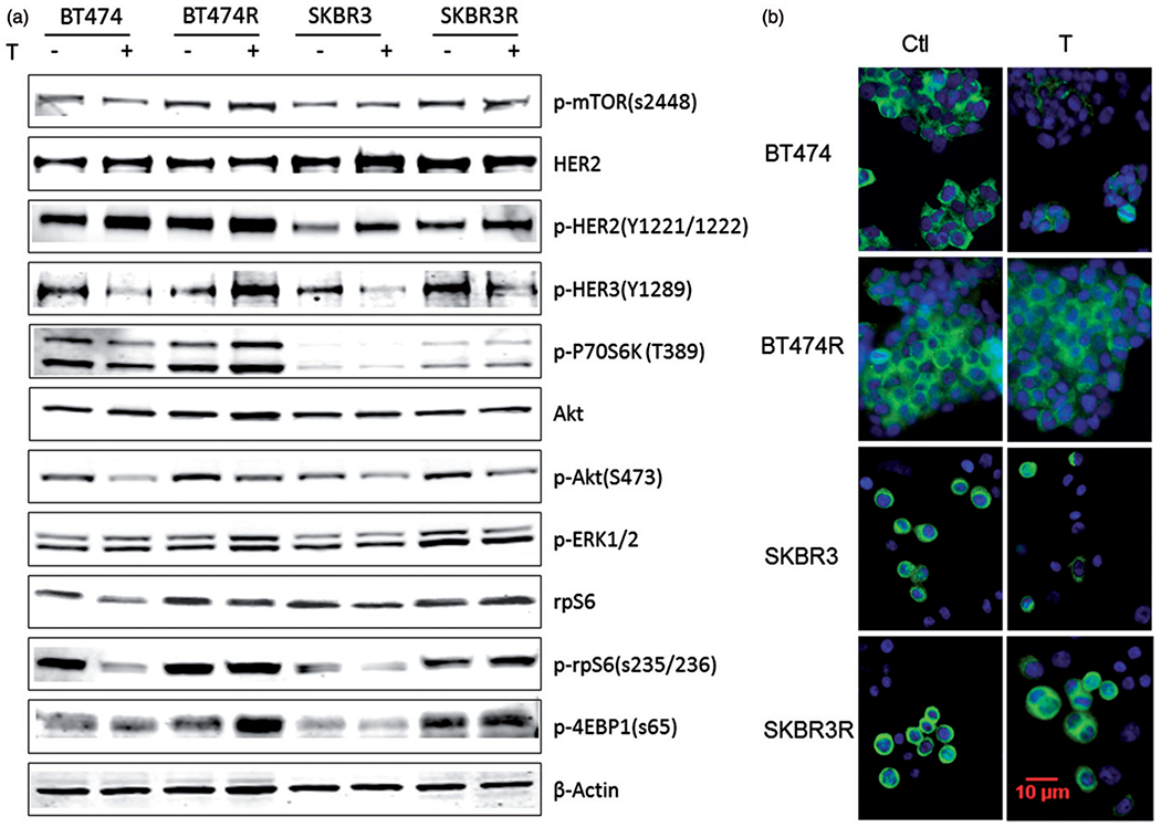
Figure 3.

p-rpS6 expression was not suppressed by trastuzumab treatment in trastuzumab-resistant BT474R and SKBR3R cells, but it was decreased in parental BT474 and SKBR3 cells. (a) Immunoblotting of whole cell lysate of key effectors in HER2 signalling pathway demonstrating altered expression of some markers in response to trastuzumab treatment. (b) Immunocytochemistry staining of p-rpS6 in BT474, SKBR3 and trastuzumab-resistant BT474R and SKBR3R cells. P-rpS6 was stained green in the cytoplasm, cell nuclei were stained with DAPI (blue lighter).
