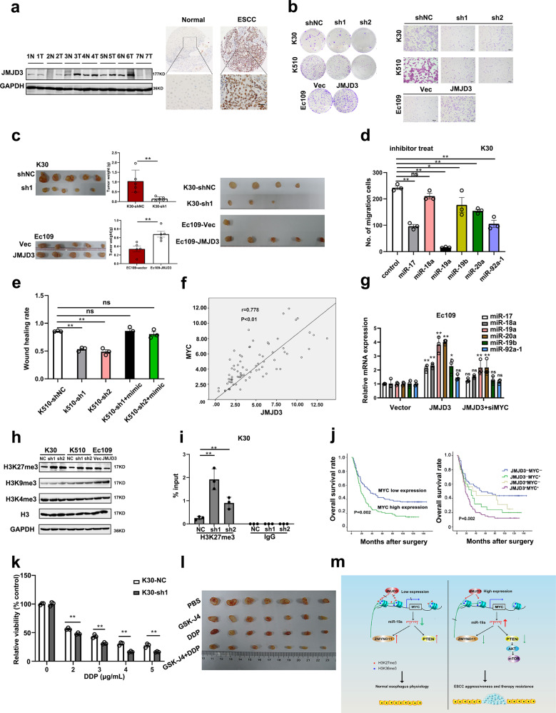
Fig. 1.
a Left panel: the protein levels of JMJD3 in seven pairs of ESCC and adjacent normal esophageal tissues. N normal tissue; T tumor tissue. Right panel: representative IHC images showing the negative expression of JMJD3 in a nonneoplastic esophageal tissue and high expression of JMJD3 in ESCC tissue. Scale bar: 50 μm. b Colony formation assay (left panel) and transwell assay (right panel) demonstrated that JMJD3 could significantly promote cell growth and migration ability in vitro. c Xenograft tumor model showed that JMJD3 could significantly promote cell growth ability in vivo (left panel), lymph node metastasis assay demonstrated that the number and size of popliteal lymph nodes decreased in JMJD3-silencing cells and increased in JMJD3 ectopic expression group (right panel). d Comparison of the effect of miR-17-92 cluster on migration using inhibitor of miRNA. e The inhibition of miR-19 could compensate the oncogenic role of JMJD3 in ESCC. f The significant positive correlation between MYC and JMJD3 was evaluated in ESCC patients from our hospital. g Silencing of MYC by siRNA in Ec109-JMJD3 cells significantly reduced the upregulation of the miR-17-92 cluster. h The abundance of H3 lysine methylation was assessed in ESCC cells by WB. i ChIP-PCR was performed to assess H3K27me3 occupancy in the MYC promoter. j Prognostic significance of MYC and JMJD3 in 161 ESCC patients assessed by Kaplan–Meier analyses. k Cell growth assay showed that JMJD3 could confer stronger chemoresistance in ESCC cell lines. l Comparison of the antitumor effects of JMJD3 inhibitor (GSK-J4) and chemotherapy (DDP) in vivo. m Graphical abstract, the upregulation of JMJD3 and its major mechanism in supporting ESCC cell aggressiveness. The results are expressed as the means ± SD of three independent experiments. [*P < 0.05, **P < 0.01, ns not significant, t-test (c, k), one-way ANOVA (d, e, g, i), Pearson correlation test (f), log-rank test (j)]

