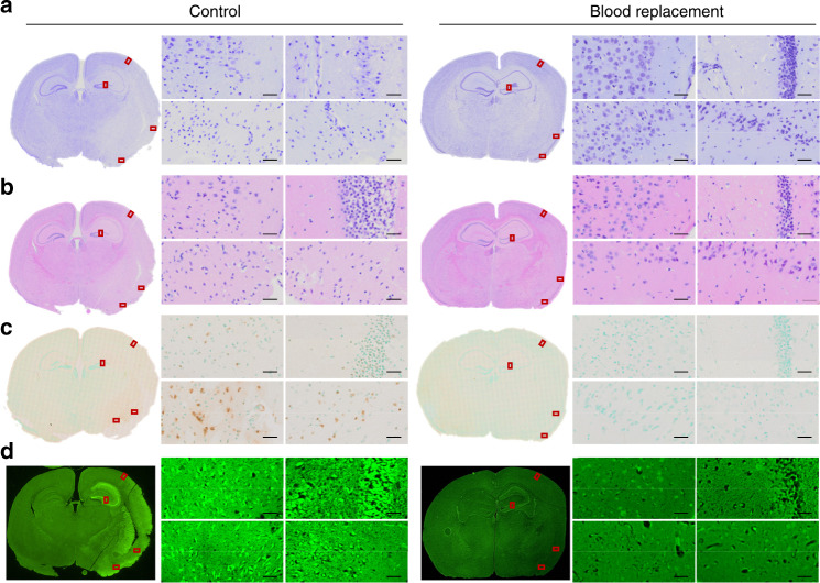
Fig. 2. Blood replacement protects the brain from cell death in stroke mice.
Microscopic images of brain tissue taken from stroke mice underwent tMCAO for 90 min followed by 22.5 h reperfusion. The blood replacement (BR) group received 500 µl of blood from healthy donors (3~6 months old) at 6.5~7 h post stroke. a The Cresyl violet staining, b H&E staining, c TUNEL staining counterstained with Methyl Green, and d Fluoro-Jade C staining were performed on paraffin embedded brain slices. Red rectangles indicate the magnified areas evaluated under ×40 magnification. Both the cortex and striatum were evaluated. Experiments were repeated five times with similar results and images were presentative of one mouse from five mice per group. Scale bars = 20 µm.

