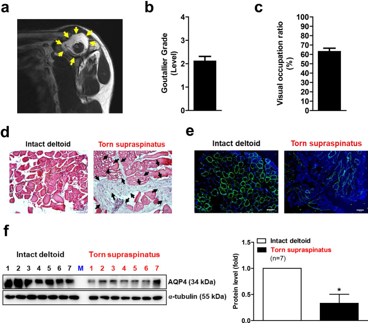
Figure 1.
Muscle atrophy and reduced AQP4 expression in the supraspinatus muscles of patients with a rotator cuff tear (RCT) (n = 9). (a) Sagittal images of postoperative MRI. Picture is a representative of independent MRI image. Yellow arrows indicate fatty infiltration in supraspinatus muscle of RCT patient. (b) The fatty infiltration of supraspinatus muscles of RCT patient was assessed using a Goutallier classification as per the following grading: grade 0, normal; grade 1, some fatty streaks; grade 2, more muscle than fat; grade 3, as much fat as muscle; and grade 4, more fat than muscle. (c) Visual occupation ratios were categorized based on a three-point scale: grade 1 involved minimal to mild atrophy of the supraspinatus muscle and an occupation ratio ≥ 60%; grade 2 involved moderate atrophy, 60% > occupation ratio ≥ 40%; and grade 3 represented severe atrophy with an occupation ratio < 40%. Age, sex, Goutallier grade, and visual occupation ratio of each patient is shown in ‘Supplementary Table 1’. (d) Histological difference between intact deltoid muscle (as a control) and supraspinatus muscle of RCT patient. Arthroscopically acquired muscle samples from the edge of the torn rotator cuff tendon were used for hematoxylin and eosin (H&E) staining. Black arrows indicate fatty infiltration within the muscle. Pictures are representative of independent tissue samples (n = 9). Scale bar: 100 μm. (e) Immunofluorescence (IF) microscopy analyses for AQP4 expression. Pictures are representative of independent tissue samples (n = 9). Scale bar: 100 μm. The negative control (secondary antibody only) for the IF showed no specific signal (data not shown). (f) Western blot analyses for AQP4 protein expression between intact deltoid muscle and supraspinatus muscle of patients with RCTs (n = 7). Densitometric analyses of the western blots are shown on the right. Data represent means ± standard errors of the means. *P < 0.05 (c).

