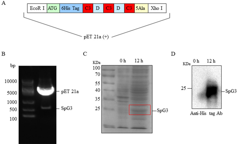
Figure 5.
The construction of pET21a-SpG3 and the expression of SpG3. (A) The structure of redesigned gene of SpG3. (B) The detection of pET21a-SpG3 by enzyme digestion. Line 1 was marker; pET21a-SpG3 in line 2 was digested with restriction enzyme EcoR I and Xho I. (C) The expression of SpG3 induced by 0.5 mM IPTG for 12 h and shaking at 30 °C. Line 1, marker; Line 2, control sample; Line 3, the sample with stimulation for 12 h. (D) Western blot. The membrane was incubated with monoclonal antibodies (mAb) against His-tag (1:3,000). Line 1, control sample; Line 2, the purified sample with stimulation for 12 h.

