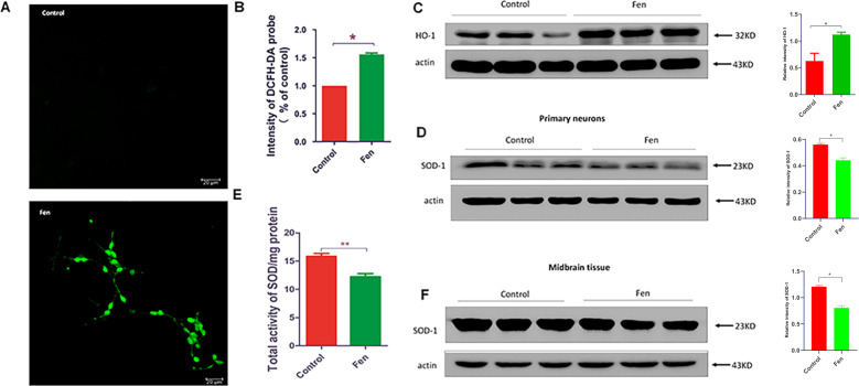
Fig. 2. Fen induces oxidative stress in primary neurons in vitro.
a Cultured primary neurons treated with Fen (100 μM) for 24 h, and intracellular ROS using the DCFH-DA probe was recorded by confocal microscopy. b Quantitative analysis of fluorescent intensity of the DCFH-DA probe by ELISA. c, d Western blotting of primary neuronal lysates was performed using antibodies to HO-1 and SOD-1 (left panel), and quantitative analysis of protein-band intensities are shown (right panel). e The activity of total SOD was measured by ELISA. f Western blotting of midbrain tissue lysates of mice was performed using antibodies to SOD-1 (left panel) and quantitative analysis of protein-band intensities are shown (right panel). For all quantitative/statistical analysis, *p < 0.05. All data are presented as means ± SEMs. Scale bars are 20 μm in a.

