Fig. 2:
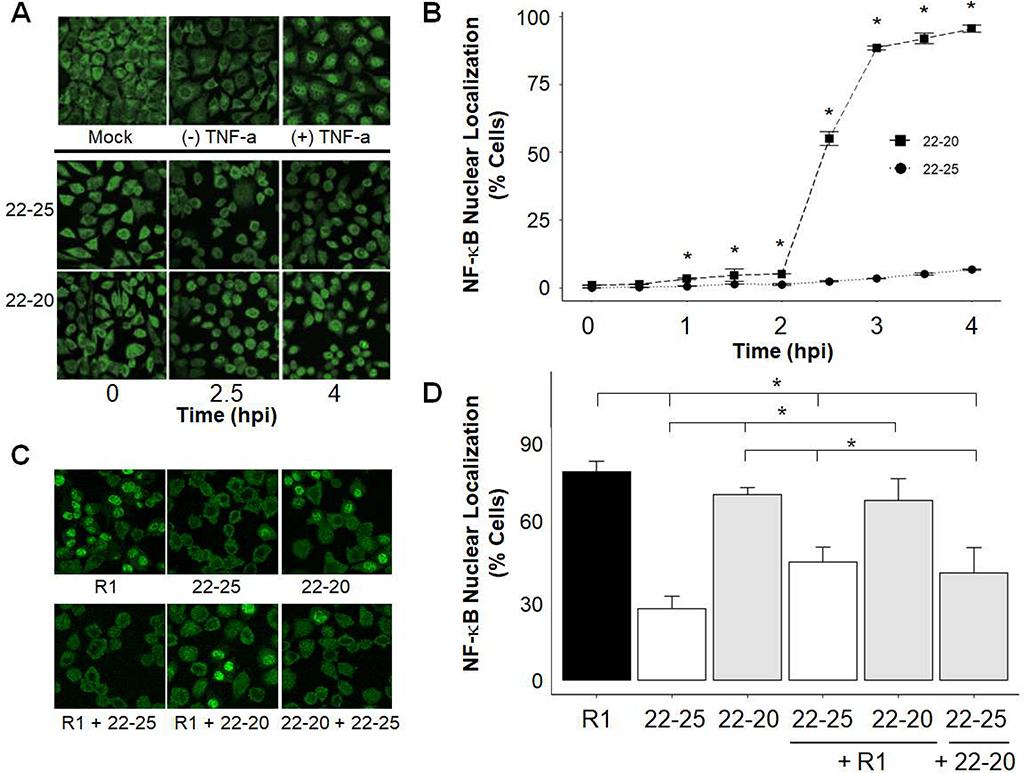
Nuclear Localization of NF-κB occurs rapidly in cells infected with viruses containing the M(D52G) and M(M51R) mutations. Cells were TNF-α treated, mock infected, or infected at an MOI of 10 for the indicated time (A, B) or coinfected at an MOI of 25 for each virus for 5 hours (C, D). The p65 subunit of NF-κB was visualized by immunofluorescence and confocal microscopy. Four or five images of each sample were taken, the total number of cells per image was counted, and the percentage of cells with nuclear NF-κB staining was determined. Data represent the mean values from three independent experiments in panel B and four independent experiments in panel D. Error bars indicate the SEM. Representative images are shown in A and C. * p-value <.05 per student’s t-test.

