Fig. 3.
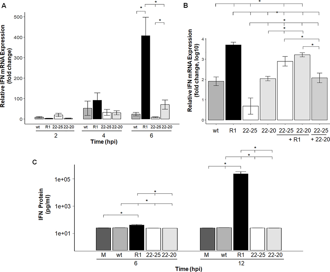
IFN-β mRNA expression and protein production are suppressed by the wt and M(D52G) strain of VSV but not by the M(M51R) virus in mouse L929 cells. L929 cells were infected at an MOI of 5 for the indicated time (A) or coinfected at an MOI of 5 for each virus for 6 hours (B). Total RNA was isolated, reverse transcribed and IFN-β mRNA quantitated by real-time PCR. Four independent experiments were performed and each sample was run in triplicate. Samples were normalized to HPRT gene expression which was stable over the time course tested. Data is represented as fold change relative to mock-infected cells. Error bar = mean +/−SEM. (C) IFN-β concentrations in VSV-infected L929 cells. Monolayers were infected with the indicated virus at an MOI of 5. At 6 and 12 hpi media from each well was collected and the concentration of IFN-β protein was quantitated by an ELISA assay. A mock sample at each time point was also collected and verified negative for IFN-β protein production. Each sample was tested in triplicate and the data shown represents the average of three separate assays. * p-value <.05 per student’s t-test.

