
Figure 4. CRISPR-Cas9 targeted APMAP gene disruption decreases JCPyV infection.
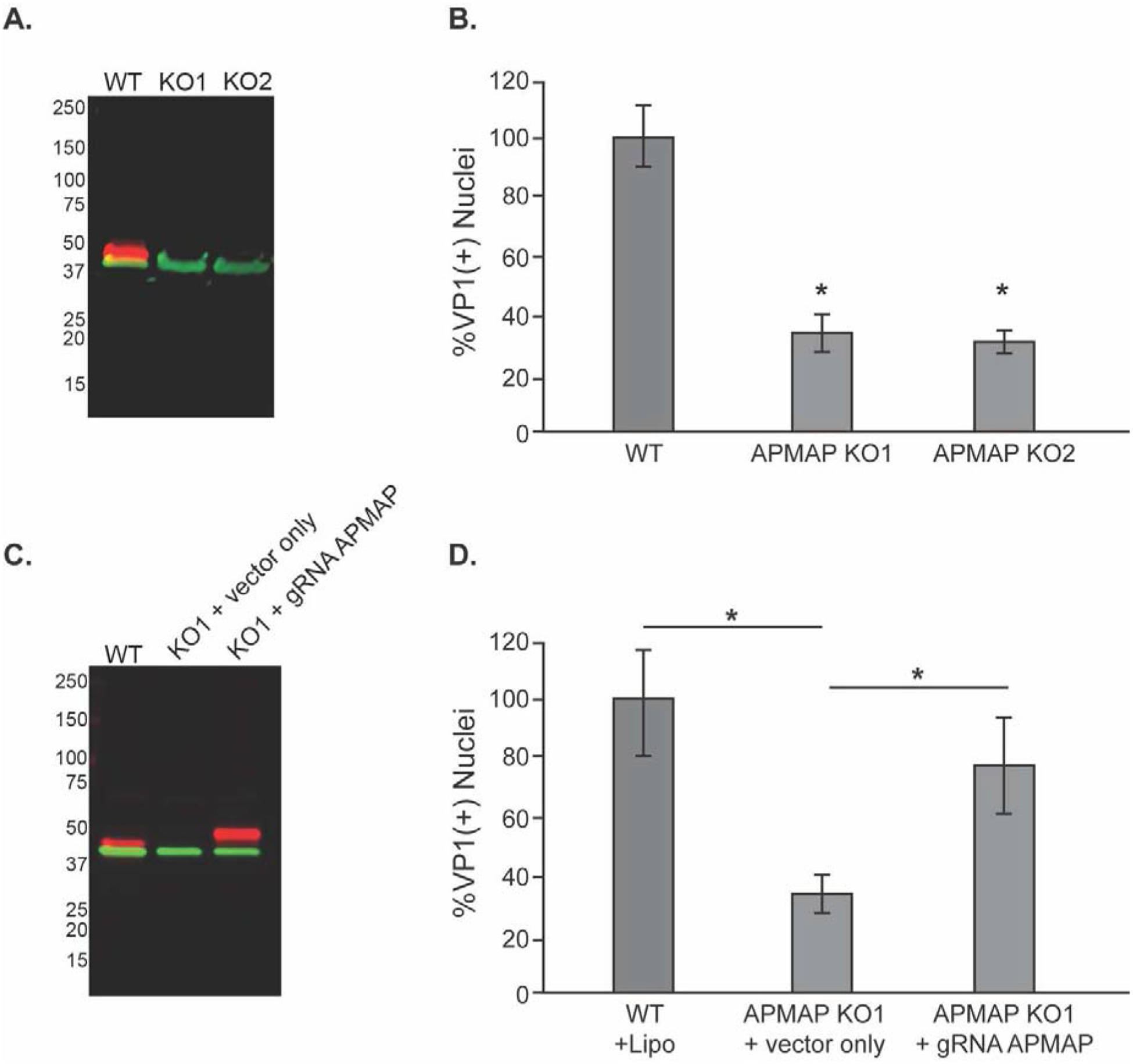
(A) Western blot analysis of wild type SVG-A (WT) cells or two APMAP null clones (KO1 & KO2) using antibodies to APMAP (labeled in red) and actin (green) demonstrates the loss of APMAP expression in the null clones. (B) SVG-A (WT) cells and APMAP null clones KO1 and KO2 were challenged with JCPyV and infection scored by indirect immunofluorescence for the viral protein VP1. (C) Western blot demonstrating APMAP expression in KO1 cells is restored by transient transfection with a plasmid expressing human APMAP with a mutated guide sequence region (gRNA APMAP). APMAP is labeled in red, actin in green as in (A). The vector-derived APMAP is slightly larger than wildtype APMAP due to a 3X flag-tag sequence from the vector that adds an additional 2.8 KDa to the C-terminal end of endogenous APMAP. (D) Susceptibility to JCPyV infection of APMAP KO1 cells was rescued by transfecting the cells with the APMAP expression plasmid expressing APMAP compared to empty control Flag expression vector without the APMAP insert. The results represent the average of three independent experiments performed in triplicate. Error bars represent SD. *p<0.05