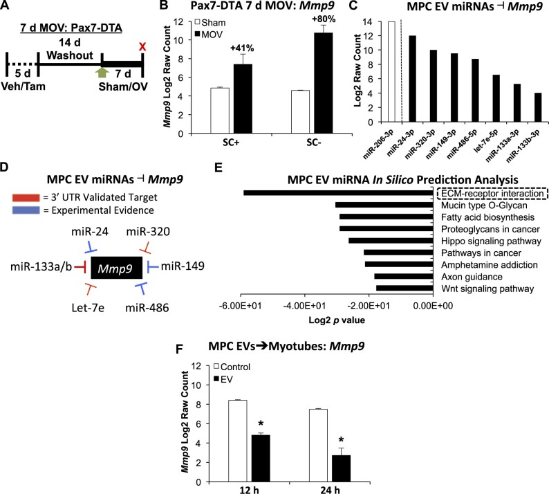
Figure 2.
Evidence for the impact of EV-mediated communication to muscle fibers in vitro and in vivo. (A) Study design schematic illustrating vehicle (Veh, SC+) and tamoxifen (Tam, SC−) treatment followed by sham or 7 days of MOV in Pax7-DTA mice. (B) Mmp9 mRNA levels in sham versus MOV in the presence and absence of satellite cells; n = 6 mice per group, two separate pools of three plantaris muscles for each group resulting in n = 2 pools per group. (C) miRNAs enriched in MPC EVs that influence Mmp9 levels in different experimental models; miR-206 was the most abundant miRNA measured. (D) Summary of evidence for miRNAs that are enriched in MPC EVs that affect Mmp9 via direct 3′-UTR targeting or indirectly via experimental manipulation using miRNA mimics and/or antagomirs (see “Results” section for specific studies). (E) DIANA miRPath analysis of miRNAs enriched in MPC EVs using the top 100 miRNAs. (F) Mmp9 mRNA levels in C57BL/6J myotubes incubated with MPC EVs for 12 or 24 h; one primary cell line was used to generate myotubes and was incubated with MPC EVs from two separate cell lines at each time point (n = 2 per time point: 12 and 24 h control, 12 and 24 h EV treatment). *Adjusted P < 0.05, all data are presented as mean ± SE.

