
Figure 5.
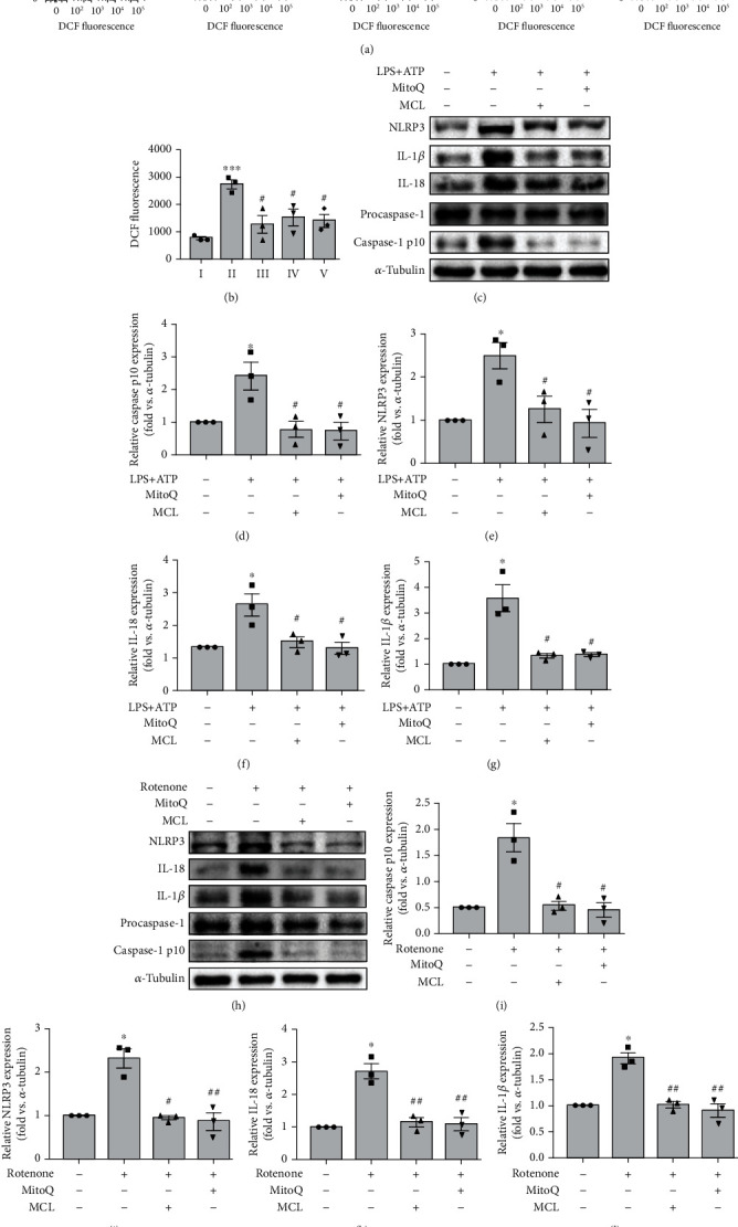
MCL inhibits NLRP3 inflammasome activation through suppressing the mROS in NRK-52E cells. (a) Flow cytometry analysis of the release of ROS in each group. (b) Comparative analysis of the amount of ROS released in each group. ∗∗∗P < 0.001 versus normal controls; #P < 0.05 versus the LPS stimulation group. (c) Western blot analysis of NLRP3, IL-1β, IL-18, and caspase-1 p10 expression in renal tubular epithelial cells treated with MitoQ (1 μM) or MCL (5 μM) and stimulated with LPS+ATP. (d–g) The relative expression levels of the indicated proteins normalized to α-tubulin expression. Data are presented as the mean ± SEM. ∗P < 0.05 versus normal controls; #P < 0.05 versus the LPS+ATP stimulation group. (h) Western blot analysis of NLRP3, IL-1β, IL-18, and caspase-1 p10 expression in renal tubular epithelial cells treated with MitoQ or MCL (5 μM) and stimulated with rotenone. (i–l) The relative expression levels of the indicated proteins normalized to α-tubulin expression. Data are presented as the mean ± SEM. ∗P < 0.05 versus normal controls; #P < 0.05, ##P < 0.01 versus the rotenone stimulation group.