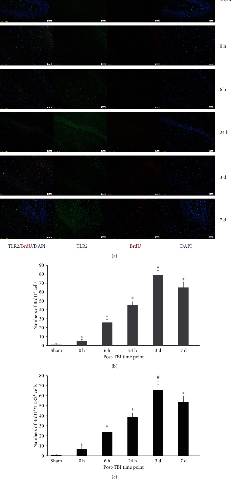
Figure 2.

Immunofluorescence (IF) microphotographs in the dentate gyrus (DG) of the DG in sham and TBI groups at different time points. (a) BrdU-labelled NSCs (red fluor), expression of TLR2 (green fluor), cell nuclei (blue fluor), and BrdU+/TLR2+/DAPI+ cells indicated that TLR2 expression in labelled proliferating cells was possible NSCs; (b) BrdU+ cells showed the proliferation of NSCs in the DG during different time points posttrauma. BrdU+ cells were more in the TBI group than that in the sham group (∗p < 0.05), and numbers of these cells were significantly different among different time points posttrauma (#p < 0.05); (c) numbers of BrdU+/TLR2+ cells indicated that the expression of TLR2 was quite different in proliferating cells of the DG among different time points posttrauma. There were more BrdU+ cells in the TBI group than that in the sham group (∗p < 0.05), and the numbers of these cells are obviously different among different time points posttrauma (#p < 0.05). Scale bar: 50 μm; data is shown as mean ± SEM.
