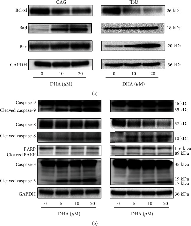
Figure 2.

DHA induces apoptosis through the extrinsic and intrinsic pathways in MM cells. (a) HMCLs were exposed to various concentrations of DHA for 24 h. The expression of the following apoptosis-related proteins was determined by western blot analysis: Bcl-xL, Bad, and Bax. (b) HMCLs were exposed to various concentrations of DHA for 24 h. The expression of the following apoptosis-related proteins was determined by western blot analysis: caspase-9, caspase-8, PARP, and caspase-3. Data are presented as the mean ± SD of at least three independent experiments.
