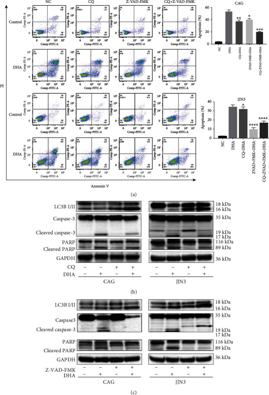
Figure 3.

The connection between DHA-induced autophagy and apoptosis. (a) HMCLs were treated with 20 μM DHA in combination with Z-VAD-FMK (100 mM), CQ (20 μM), or both, and apoptosis was evaluated after 24 h by flow cytometry. The data are presented as the mean ± SD of three independent experiments. (b) HMCLs were preincubated with CQ (20 μM) for 2 h, followed by treatment with the indicated concentrations of DHA for an additional 24 h. Whole-cell lysates were prepared for western blot analysis of LC3B, caspase-3, and PARP. (c) HMCLs were preincubated with Z-VAD-FMK (100 mM) for 2 h, followed by treatment with the indicated concentrations of DHA for an additional 24 h. Whole-cell lysates were prepared for western blot analysis of LC3B, caspase-3, and PARP. Data are presented as the mean ± SD of at least three independent experiments. ∗P < .05, ∗∗P < .01, ∗∗∗P < .001, ∗∗∗∗P < .0001.
