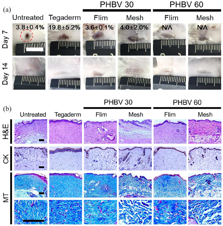
Figure 5.
Wound regeneration in a full-thickness in vivo mouse wound model. Wounds were treated with PHBV 30 and PHBV 60 scaffolds (film or mesh), or with TegadermTM (positive control). (a) Gross appearance of wounds, indicating open wound areas (%) at days 7 and 14 after wound dressing (scale bar, 1 cm). (b) Histological analysis of regenerated wounds at day 28, staining with H&E, anti-cytokeratin antibody (CK), and Masson’s trichrome (MT) (scale bar, 100 µm).
*p < 0.05 by one-way ANOVA.

