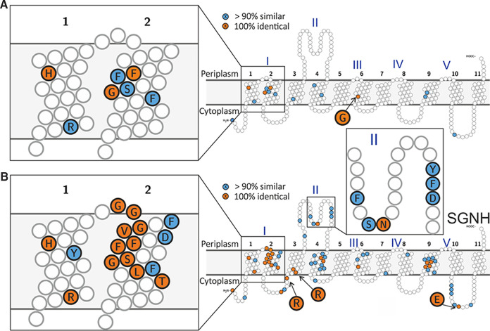
FIG 2.
Conservation in transmembrane domains of experimentally characterized bacterial AT3 carbohydrate acetyltransferases. The 100% identical residues are colored orange, similar residues in >90% sequences are colored blue, and conserved small hydrophobic residues in transmembrane helices were not colored. (A) Conserved residues across all 30 currently known experimentally characterized proteins and OafB-SPA. (B) Conservation in only AT3-SGNH fused proteins in the alignment. See Table S1 for details of aligned sequences and Fig. S1 for full alignment.

