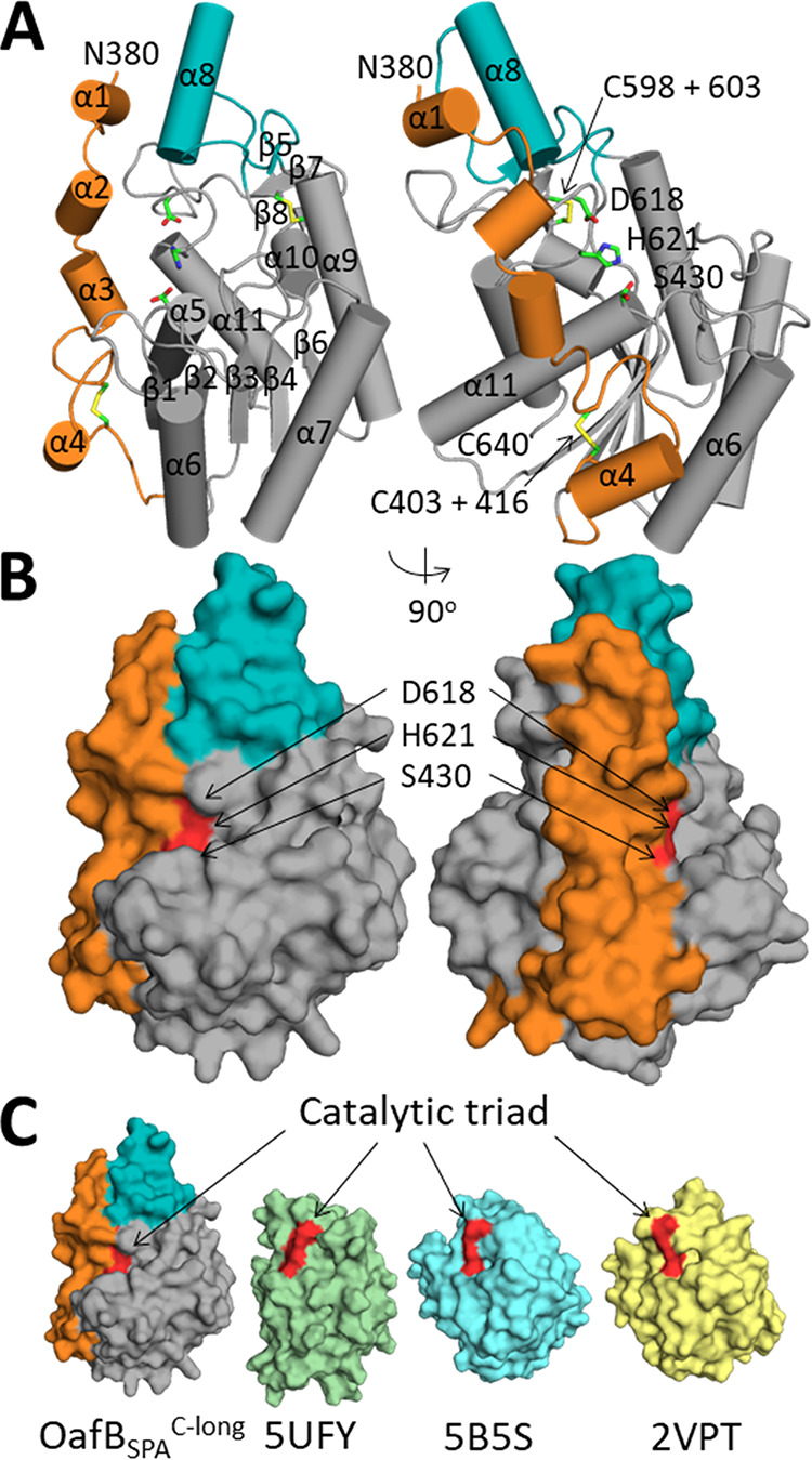
FIG 5.

Analysis of the crystal structure of OafB-SPAC-long. (A) Cartoon representation of OafB-SPAC-long with helices and sheets numbered, with the additional helix (α8) colored teal and SGNH-extension colored orange. Catalytic residues and disulfide bonds are shown as sticks and are labeled. (B) Surface representation of OafB-SPAC-long with coloring as above and the catalytic triad colored red. (C) Surface representation of OafB-SPAlong, PDB 5UFY, PDB 5B5S, and PDB 2VPT.
