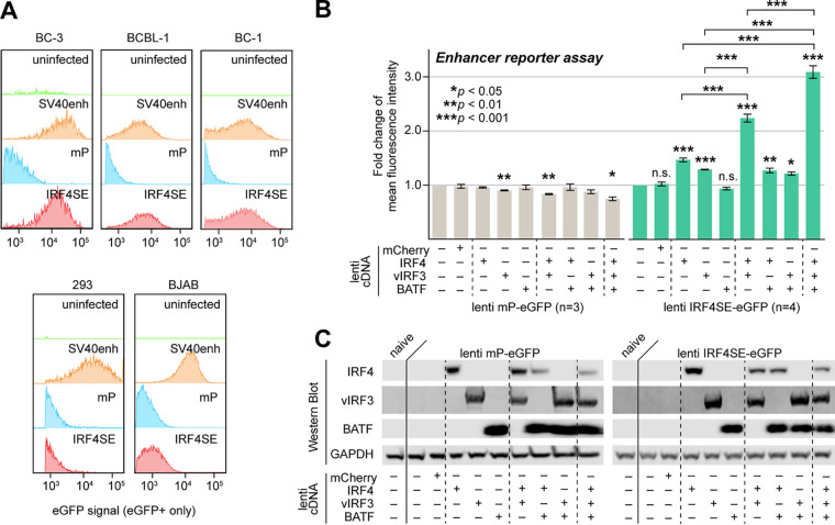
FIG 5.
IRF4, vIRF3, and BATF cooperatively promote IRF4-SE activity. (A) A 500-bp sequence centered on the prominent IRF4 and vIRF3 ChIP-Seq peaks ∼63 kb upstream of the IRF4 TSS drove eGFP expression from a lentiviral enhancer reporter (IRF4SE-eGFP) after transduction into PEL cell lines BC-3, BCBL-1, and BC-1 but not the IRF4/vIRF3-negative cell lines 293 and BJAB. Cell lines were transduced at MOI 3 and analyzed 4 days after transduction. Similar reporters containing the SV40 enhancer or a minimal promoter (mP) served as positive or negative controls, respectively. Reporters were titrated by qRT-PCR and, where possible, FACS analysis prior to transduction. Data are representative of results from n = 3 biological replicates. (B) The lentiviral IRF4SE or mP eGFP reporters were transduced into 293 cells at MOI 3, together with the indicated combinations of lentiviral expression vectors for vIRF3, IRF4, or BATF. Expression of mCherry served as a negative control. eGFP mean fluorescence intensities were measured by FACS analysis on day 3 after transduction and are shown relative to values from cells that were not transduced with lentiviral cDNA expression vectors. Error bars represent SEM from 3 (mP) or 4 (IRF4SE) biological replicates. *, P < 0.05; **, P < 0.01; ***, P < 0.001 (from paired two-sided Student's t tests). n.s., not significant. (C) Western blots from experiments performed as described for panel B, demonstrating ectopic expression of IRF4, vIRF3, and/or BATF as indicated. The blots shown are representative of n = 3.

