
Figure 3. Differential cortical feedback bouton odor responses across bulb layers.
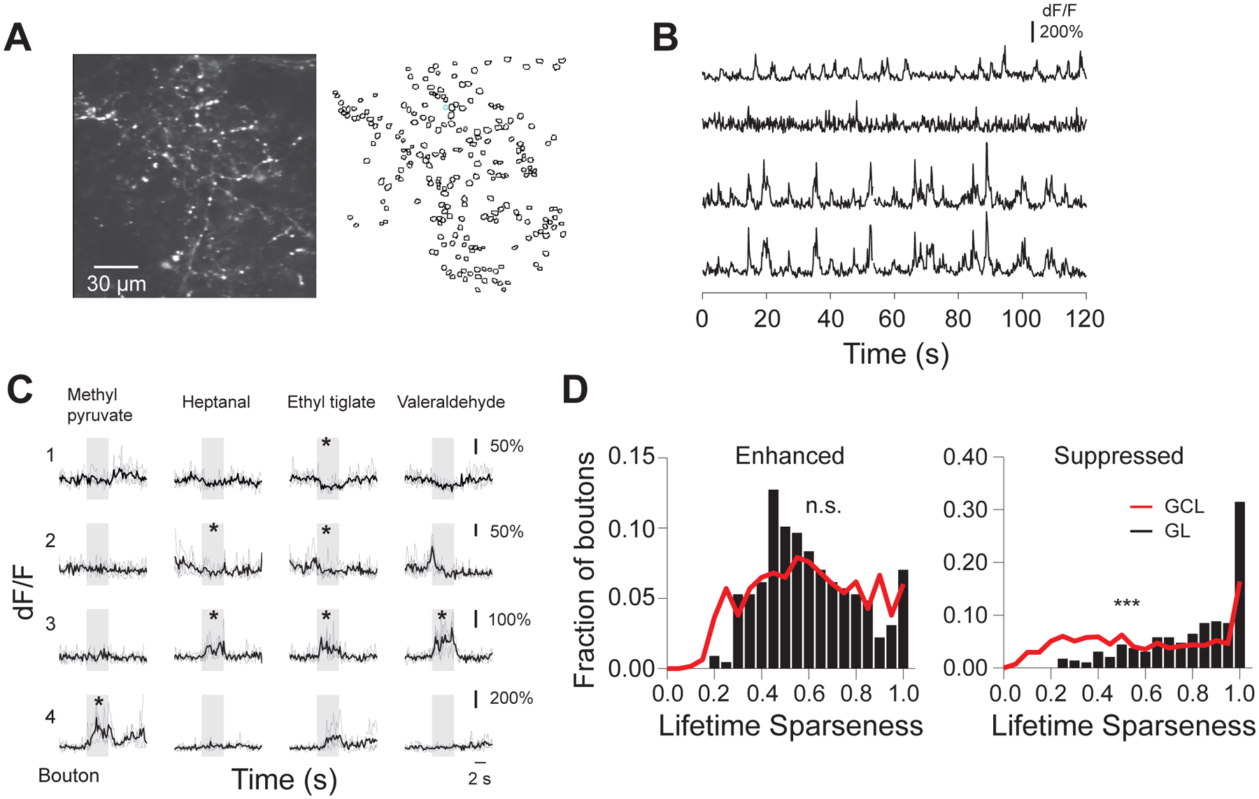
A. (Left) Example field of view ~80 μm deep from surface of GCaMP5 labeled cortical feedback axons and boutons in an awake head-fixed mouse; (Right) Outlines of the regions of interest (ROIs) corresponding to putative cortical feedback boutons;
B. Spontaneous activity traces from the feedback boutons selected in the field of view showed in A. Bottom two traces correspond to boutons assigned to the same axonal branch by reconstruction of single axons;
C. Odor responses of four example boutons in GL across four different stimuli (ethyl pyruvate, heptanal, ethyl tiglate, valeraldehyde, 0.4% saturated vapor pressure). Individual repeats (gray) and average traces (black) are shown; odors trigger both positive (enhanced responses) and negative (suppressed responses) deflections from baseline; * mark significant odor responses; stimulus duration, 4s;
D. Lifetime sparseness of boutons responsive to odors in the panel (Odor Set A, Table S1) only via enhancement (Left) or suppression (Right); distributions in the GCL (black bars) and GL (gray trace) are shown.