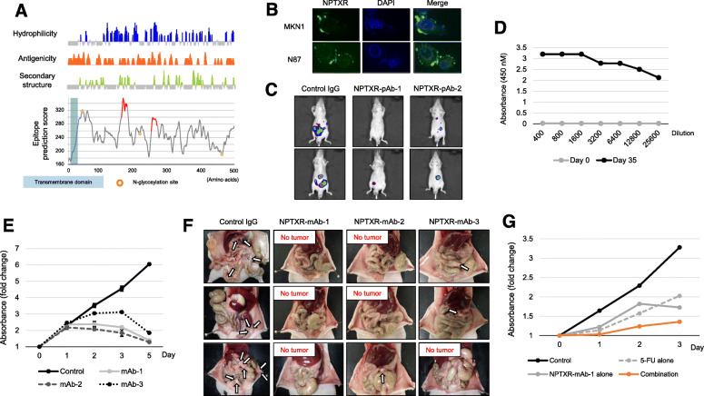
Fig. 4.
Characterization of anti-NPTXR pAbs and mAbs. a Bioinformatics analysis of predicted immunogenic epitopes in NPTXR. b Immunofluorescence microscopy of MKN1 and N87 cells labeled with NPTXR-pAb-1 and Alexa Fluor 488 (green). Nuclei were stained with DAPI (blue). c IVIS analysis of representative BALB/c nu/nu mice injected with luciferase-expressing parental or NPTXR-KO MKN1 cells. Mice were injected with d-luciferin for 15 min and imaged 3 weeks after cell injection. d ELISA assay of sera from mice 3 weeks after immunization and boosting with NPTXR peptide. e CCK-8 proliferation assay of MKN1 cells incubated with NPTXR mAb-1, − 2 and − 3. f Therapeutic effects of intraperitoneal administration of anti-NPTXR monoclonal antibodies. g CCK-8 proliferation assay of MKN1 cells treated with 5-fluorouracil (5-FU) and NPTXR-mAb-1. *P < 0.05. Mean ± standard deviation

