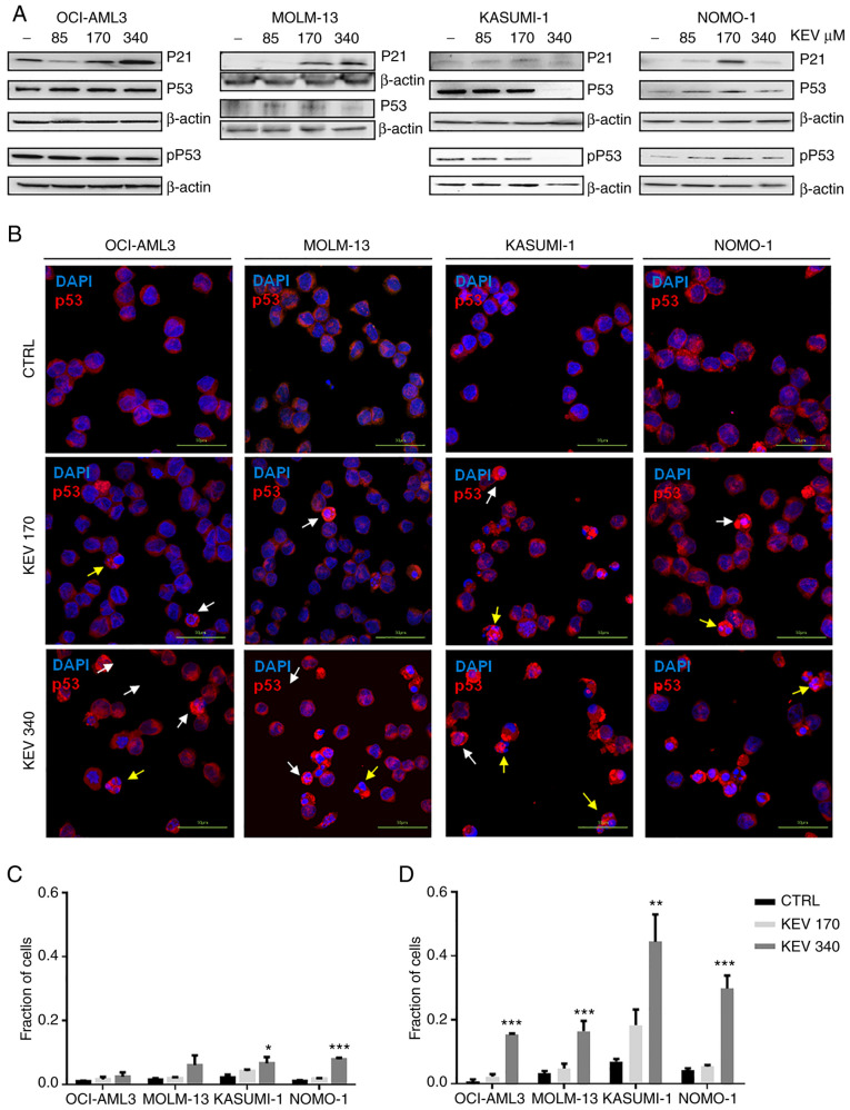
Figure 5.
Effect of kevetrin on the expression of p53 in AML cell lines. (A) Representative western blots of pSer15 p53 (pP53), P53 and P21 expression levels in MOLM-13, KASUMI-1, OCI-AML3 and NOMO-1 cell lines treated with kevetrin at 85, 170 and 340 µM for 48 h. β-actin was used as loading control. The figures show one representative of three independent experiments. (B) Immunofluorescence analysis of p53 in AML cell lines treated with 170 and 340 µM kevetrin for 48 h. The nuclei were stained with DAPI. White and yellow arrows indicate intact cells with nuclear p53 and nuclear-fragmented cells expressing high p53, respectively. (C) Fraction of intact cells with nuclear p53 localization or (D) p53-high apoptotic cells in kevetrin-treated cells. Values represent the mean ± standard deviation of 3 biological replicates (*P<0.05, **P<0.01, ***P<0.001). AML, acute myeloid leukemia; KEV, kevetrin; CTRL, control.

