Fig. 1.
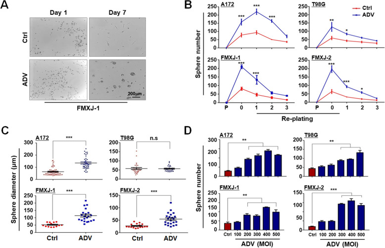
ADV infection promotes tumor sphere formation by glioma cells. a Primary GBM cells (FMXJ-1) were infected with ADV for 8 h, and then cultured under the neurosphere condition for 7 days and photographed. b Primary and lined glioma cells (P) cultured under ordinary condition without the sphere supplements). Cells were infected and cultured as in (A) for 7 days (re-plating 0). Spheres were then re-plated serially every 7 days for 3 times (as re-plating generation 1, 2, and 3, respectively). Number of tumor spheres was counted on each generation. Cell not infected with ADV were used as controls. c Diameter of spheres on day 7 was measured. d Primary and lined glioma cells were infected with different amounts of ADV (MOI) and cultured under the neurosphere condition for 7 days. Number of tumor spheres was counted. Data are represented as mean ± SEM, n = 6. *, P < 0.05; **, P < 0.01; ***, P < 0.001; n.s, not significant

