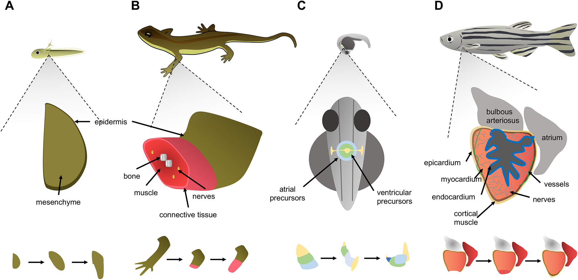
Figure 1. A comparison between patterning in embryogenesis and regeneration events.

A. Growth of forelimb buds in the larval newt begins with an early bud stage before skeletal elements form, indicating a simple developmental primordium that grows and becomes patterned as the entire organism grows. Amputation stimulates the formation of a primordium called a blastema from remaining cells, which similar to a limb bud, grows and is patterned into mature skeletal elements, connective tissue, glands, nerves, skeletal muscle and epidermis. B. During zebrafish embryogenesis, mesoderm-derived cells destined to form the cardiac atrium and ventricle are arranged in a ring or cone pattern, prior to patterning into chambers and acquiring contractile activity. An adult zebrafish ventricle is comprised of multiple types of innervated and vascularized heart muscle surrounded by an outer layer of epicardial cells and coated internally by a layer of endocardial cells. After resection of the ventricular apex, spared cardiomyocytes divide and non-muscle tissues are re-established. Injury is indicated by a lightning bolt icon.
