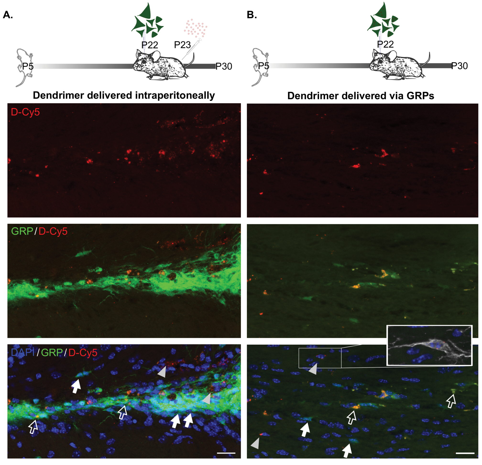
Figure 2:

A) Representative images of corpus callosum in mice receiving D-Cy5 24 hr after P22 GRP transplantation. D-Cy5 is detected within GRPs (white open arrows) and outside of GRPs (gray arrowheads). GRPs without dendrimer are indicated by a white filled arrow. B) Dendrimer was similarly detected both inside of GRPs (white open arrows) and outside of GRPs (gray arrowheads), with D-Cy5 negative GRPs also present in the area (white filled arrows). Dendrimer is also detected within other cell types as indicated by inset showing Iba1+ microglial cell. Scale bars equal 20 μm.
