
Figure 13.
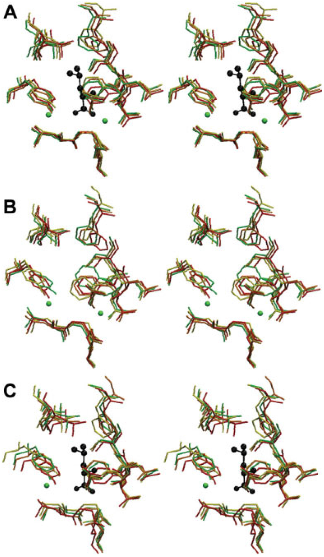
A stereo image of LeuT binding pocket at different intervals of MD simulation. Residues pictured contain at least one atom crystallized within 4.50Å of a nonhydrogen atom found in the coordinates for the crystallized leucine substrate (black). Clockwise from top, the LeuT binding pocket is defined by F259, S256, L255, T254, F253, A22, N27, G26, L25, Y108, and I359. The binding pocket is pictured at 0 (red), 10 (orange), 15 (yellow), and 20 (green) nanoseconds of simulation time. A: MD simulation in the presence of substrate. B: MD simulation in the absence of substrate. C: MD simulation in the presence of substrate but without the sodium ion crystallized closest to the carboxyl group of the substrate.