Fig. 6.
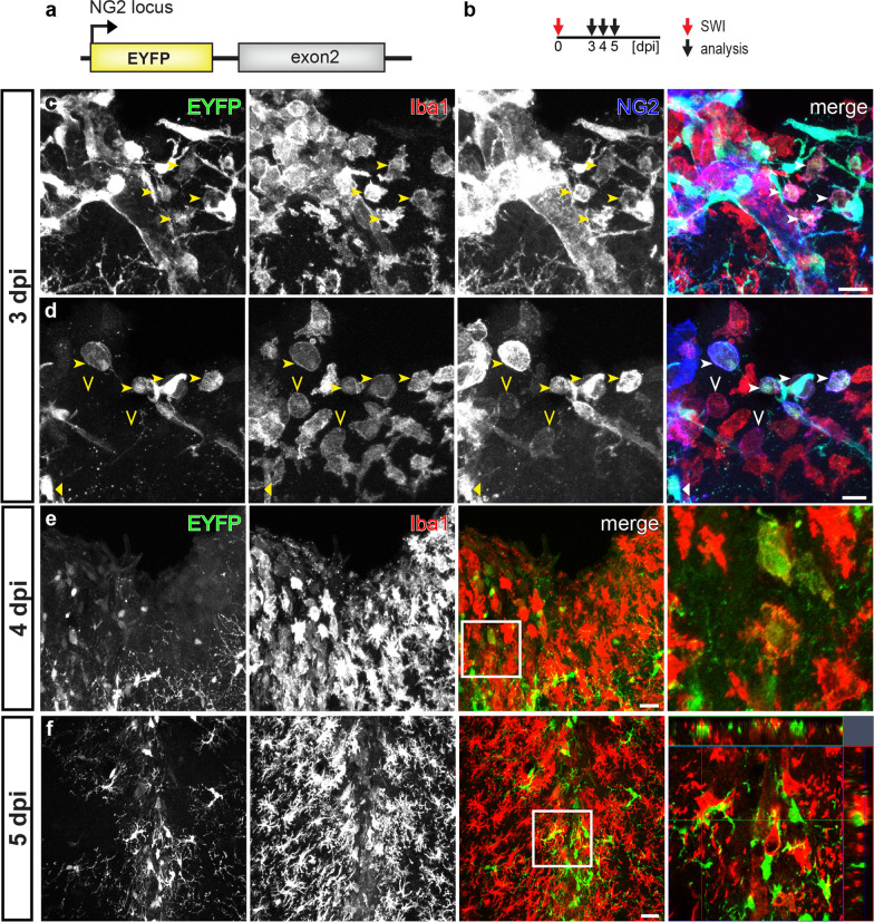
Few microglia in NG2-EYFP mice with NG2 promotor activity after SWI. a Schematic of transgene structure of NG2-EYFP mice used in SWI model. b Experimental plan: NG2-EYFP mice were analyzed at 3, 4, or 5 dpi (SWI). c and d Confocal micrographs showing EYFP+, Iba1+, and NG2+ cells at the lesion site at 3 dpi. A few NG2+ Iba1+ cells could be detected at the lesion site with (arrowheads in c and d) or without (open arrowheads in d) expression of EYFP. e and f Confocal images showing EYFP and Iba1 immuno-positive cells at the injured area at 4 (e) and 5 (f) dpi. EYFP+ Iba1+ cells could be rarely observed at 4 dpi (e, white box showing an example magnified in the right image), and were almost undetectable at 5 dpi (g, white box was shown as orthogonal view in the right image). Scale bars = 20 μm

