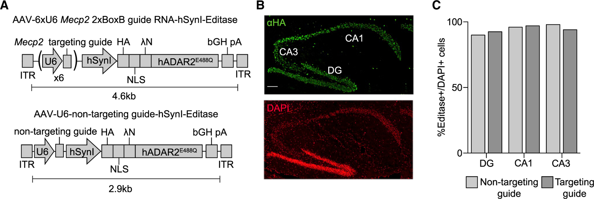
Figure 1. Hippocampal Expression of RNA Editing Components following Stereotaxic Injection of Mecp2317G>A Mice.

(A) Schematic of AAV editase expression vectors. Each construct contains the human Synapsin I promoter for neuronal editase expression and either six individual U6 promoters, each driving expression of one copy of the Mecp2 2xBoxB targeting guide (top) or a single human U6 promoter driving expression of a small non-targeting RNA (bottom).
(B) Confocal images of a Mecp2317G>A mouse 3 weeks after hippocampal injection of the AAV PHP.B vector. HA immunostaining identifies the editase in the dentate gyrus (DG) and CA1 and CA3 pyramidal neuronal layers. Scale bar, 100 μm.
(C) Quantification of HA-editase-positive cells for each virus relative to the total number of cells in each region (mean, n = 2 mice per condition). More than 100 cells were counted per hippocampal region per replicate.
