
Figure 2. Efficient Editing of Mecp2 RNA following Hippocampal Injection of Mecp2317G>A Male Mice (Post-natal Day 28 [P28]).
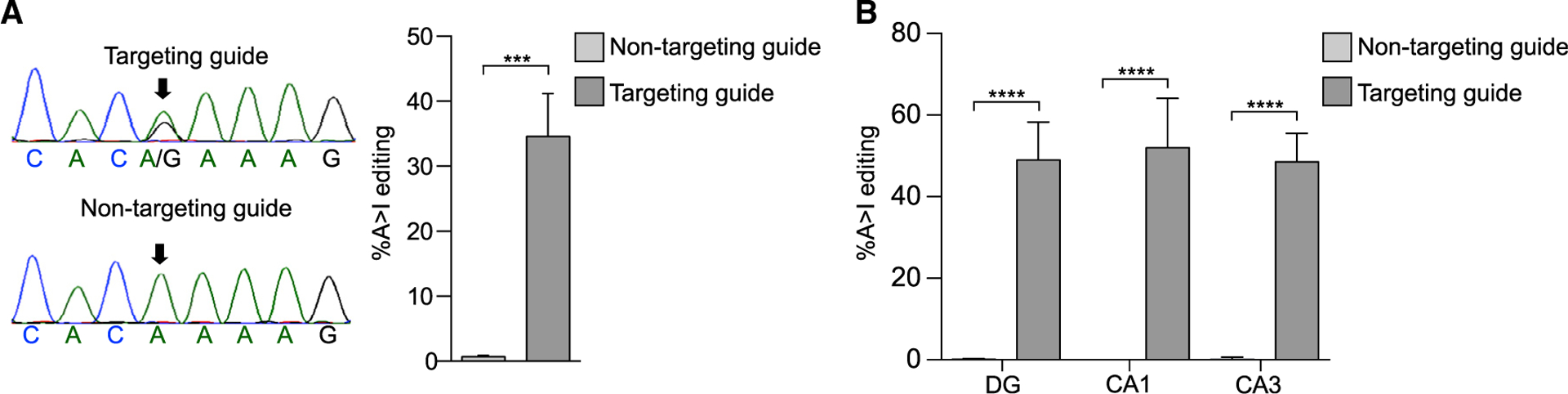
(A) Left: sequencing chromatograms of cDNA from an intact hippocampus injected with the editase and the indicated guides 3 weeks after viral injection. An arrow denotes the on-target base. Right: quantification of editing (mean ± SD, n = 3 mice per condition). ***p < 0.01 unpaired two-tailed t test.
(B) Quantification of editing in hippocampal neurons following laser capture micro-dissection. Mean ± SD, n = 3 mice/condition 3 weeks after viral injection. ****p < 0.001, one-way ANOVA and Tukey’s multiple comparisons test.