FIG 4.
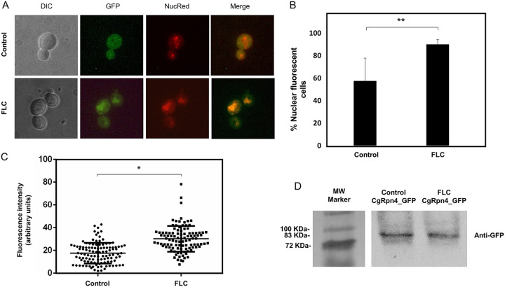
CgRpn4 increases its relative distribution to the nucleus upon fluconazole stress. The subcellular localization of fluorescence in exponential-phase L5U1 C. glabrata cells harboring the pGREG576_MTI_CgRPN4 plasmid after 5 h of recombinant protein production under control conditions or after 1 h of fluconazole exposure was assessed. (A) Representative images of CgRpn4_GFP localization in L5U1 C. glabrata cells. DIC, differential interference contrast; GFP, green fluorescent protein; NucRed, nuclear stain; merge, GFP-NucRed overlap images. (B) Percentage of cells showing nuclear localization of the CgRpn4_GFP fusion protein under control conditions or after 1 h of fluconazole exposure. Error bars represent the corresponding standard deviations. *, P < 0.01. (C) Comparison of nuclear fluorescence intensities of CgRpn4_GFP in L5U1 C. glabrata cells under control conditions or after 1 h of fluconazole exposure. The estimation of nuclear signal intensity was calculated after deduction of the cytoplasm intensity and correction for background intensity. Error bars represent the corresponding standard deviations. *, P < 0.05. (D) Western blot detection of the CgRpn4_GFP fusion protein under control conditions or after 1 h of fluconazole exposure. Immunoblotting was carried out using a mouse anti-GFP antibody. The displayed images are representative of results from two independent experiments. MW, molecular weight.

