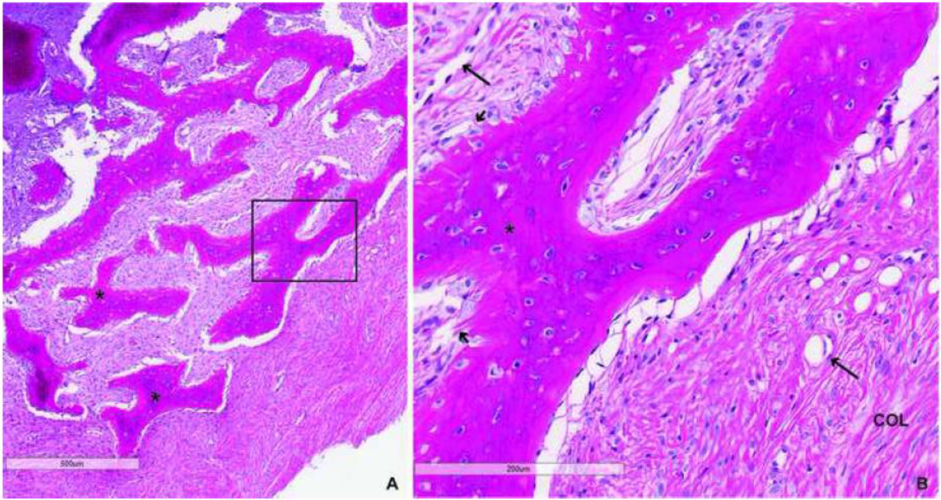
Figure 3.

H&E staining of fibrous dysplasia (FD) biopsy: Low (A) and high power (B) images demonstrate typical features of FD including abnormal, hyperosteocytic bony trabeculae (*) with classic “Chinese-writing” appearance, cellular fibrotic stroma with prominence of vascular structures (long arrows), Sharpey fibers (short arrows), abundant collagen fibers (col) and an absence of hematopoietic marrow or adipocytes. Importantly, there was no histological evidence of malignancy.
