Figure 2.
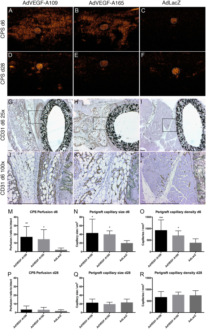
Perfusion ultrasound and perigraft capillaries. Perfusion ultrasound (A, B, C, D, E and F) and immunohistological staining for endothelial cells (G, H, I, J, K and L). CPS ultrasound performed 6 days after gene transfer shows greatly increased perfusion surrounding the graft after treatment with AdVEGF-As (A and B) with contrast agent visible mainly inside the graft lumen in controls (C). At day 28, the perfusion increase has attenuated and a strong signal is only acquired from patent graft lumens (D, E and F). CD31 immunohistology shows an increase in the number and size of adventitial capillaries surrounding the grafts with both AdVEGF-A treatments (G, H, J and K). At day 6, both VEGF-A treatments increased perfusion (M) as well as the size (N) and number (O) of adventitial capillaries. These effects were transient and had dissipated by day 28 (P, Q and R). Graft lumen outlined with dotted white line in A, B, C, D, E, F. Box in G, H, I indicates area for higher magnification in J, K, L. Asterisks in M, N, O, P, Q, R indicate significant P values with *P < 0.05, **P < 0.005, ***P < 0.001. Scale bar 500 µm in G, H, I, J, K, L.

 This work is licensed under a