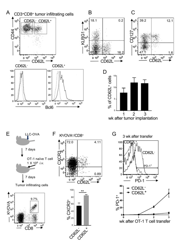
Fig 1. Memory phenotype CD8 T cells expressing Bcl6 persist within the tumor.
(A–C) C57BL/6 mice were intradermally transplanted with LLC-OVA. Representative flow cytometry plots and histograms of tumor-infiltrating CD3+CD8+T cells on day 7 of three independent experiments are shown. (A) Bcl6 expression in sorted CD62L+ and CD62L- cells. (D) Frequencies of CD62L+ cells within the CD3+CD8+ tumor-infiltrating cells. Representative data of three independent experiments are shown. (Mean ± SEM, n = 3) (E) Experimental design. OT-1 naïve T cells were adoptively transferred into C57BL/6 mice that had been transplanted with LLC-OVA 7 days previously. Kb/OVA tetramer+ cells were analyzed on day 7. (F) Flow cytometric analysis of Kb/OVA+ cells and frequencies of CXCR3+ cells. Representative data of three independent experiments are shown. (Mean ± SEM, P ** <0.01, n = 3) (G) PD-1 levels of CD62L+ and CD62L- tumor-infiltrating OT-1 T cells 3 weeks after the transfer. A representative analysis of three independent experiments is shown (top). Frequencies of PD-1+ cells in CD62L+ and CD62L- populations 1–3 weeks after OT-1 T cell transfer (bottom). Data are shown as mean ± SEM (n = 3). Representative data of three independent experiments.

