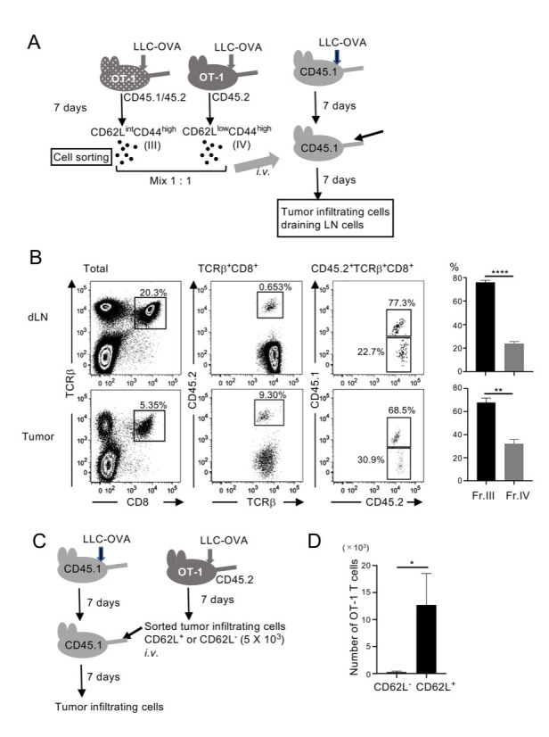
Fig 3. CD62LintCD44high T cells both in tumor-draining lymph nodes and within the tumor have a strong potential to proliferate.

(A) Experimental design. Kb/OVA+CD62LintCD44high (III) and CD62L-CD44high (IV) cells in tumor-draining lymph nodes in LLC-OVA–transplanted OT-1(CD45.1/45.2) and OT-1(CD45.2), respectively, were sorted. Cells were mixed at a 1:1 ratio and transferred i.v. into LLC-OVA–transplanted wild-type (CD45.1) mice. Transferred OT-1 T cells in the tumor and tumor-draining lymph nodes were analyzed on day 7. (B) Representative flow cytometry plots of two independent experiments. Frequencies of CD45.1+ cells (from fr.III) and CD45.1- cells (from fr.IV) among TCRβ+CD8+ CD45.2+ cells were analyzed. Mean ± SEM; **** P<0.001, ** P<0.01; n = 3. (C) Experimental design. Kb/OVA+CD62L+ and CD62L- tumor-infiltrating cells were sorted from LLC-OVA–transplanted OVA mice. Cells (5 × 10³) were transferred into LLC-OVA–transplanted wild-type (CD45.1) mice. Tumor-infiltrating cells were analyzed on day 7. (D) Numbers of cells derived from transferred OT-1 T cells within the tumor were calculated from the cell numbers passed through the FACS and their profiles. Data shown are representative of two independent experiments. Mean ± SEM; * P<0.05; n = 3.
