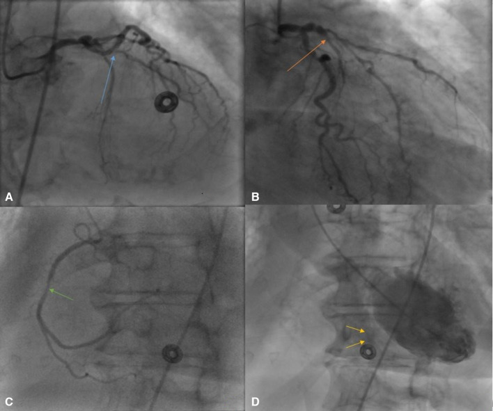
Figure 5.
(A) Coronary angiogram (CA) in left anterior oblique (LAO) cranial view shows total occlusion of proximal left anterior descending artery (LAD) (blue arrow). (B) CA in right anterior oblique (RAO) caudal view shows occlusion of proximal LAD (orange arrow). (C) CA in LAO cranial view shows 40% stenosis of right coronary artery (green arrow). (D) Left ventriculogram in LAO cranial view shows left to right shunt with dye in right ventricle (yellow arrows).

