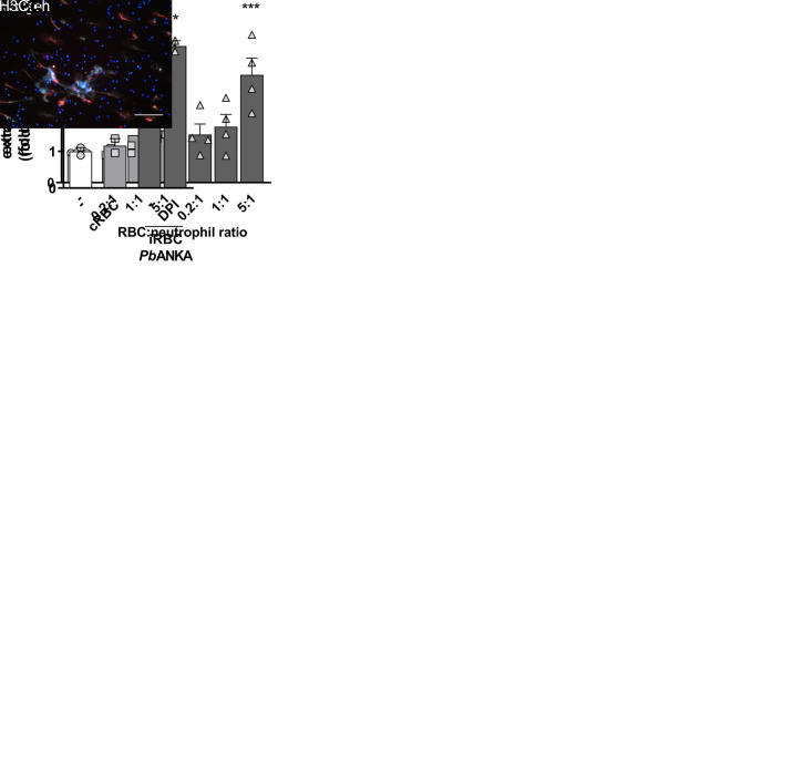
Fig 6. P. berguei ANKA-infected erythrocytes induce NETs.
(A) Fluorimetric determination of NET production by murine neutrophils in the presence of P. berguei ANKA-infected mouse red blood cells (iRBC PbANKA) or uninfected RBC (cRBC) at varying red blood cell:neutrophil ratios. (B) Mouse neutrophils were pre-treated for 30 minutes with DPI and then incubated with PbANKA-infected RBC. NET production was determined by fluorimetry as before. Data in A and B are presented as means ± S.E.M. of the fold induction of extracellular DNA signal relative to resting neutrophils. (C) Visualization by fluorescence microscopy of NETs produced by murine neutrophils in the presence of PbANKA-infected (iRBC) or uninfected (cRBC) red blood cells. DNA is stained in blue (Hoesch), myeloperoxidase is stained in green (MPO) and citrullinated histone H3 (H3C) is stained in red. Data are presented as means ± S.E.M. of the percentage of infected red blood cells. * P< 0.05 and ** P<0.01 relative to untreated controls.

