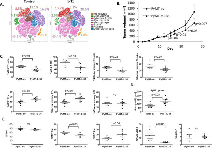
Figure 1.
IL-31 expression in tumors enhances antitumor immunity. (A) Non-tumor-bearing mice were infused with IL-31 via osmotic pumps (~14 µg/day) or control (n=5 mice/group). Three weeks later, mice were sacrificed and spleens were removed. Splenocytes (pooled per group) were immunostained with a 37-antibody panel as described in online supplementary table S1. Cells were acquired by CyTOF and data were analyzed by Cytobank. The analysis of the different immune cell populations found in the spleens is presented by viSNE plot. (B–D) PyMT cells stably overexpressing IL-31 (PyMT-IL-31) or their counterpart control cells (PyMT-ev) were implanted in the mammary fat pads of 10-week-old C57BL/6 female mice (n=6 mice/group). Tumor volume was measured and plotted (B). At endpoint, tumors were removed and prepared as single cell suspensions. Lymphoid cells (C), Granzyme B expression in tumor lysates (D), and myeloid cells (E) were assessed. Statistical significance was assessed by unpaired two-tailed t-test. Significant p values are shown. CTL, cytotoxic T lymphocyte; CyTOF, time of flight mass cytometry; IL-31, interleukin-31; MDSCs, myeloid-derived suppressor cells; NK, natural killer; ns, non-significant; PMN, polymorphonuclear; TAM, tumor-associated macrophage; Th, T helper.

