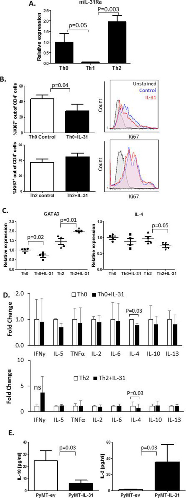
Figure 2.

IL-31 affects CD4+ T cell proliferation and cytokine secretion. (A–D) CD4+ T cells were isolated from spleens of naïve C57BL/6 female mice by immunomagnetic negative selection. The cells were left untreated (Th0) or skewed toward Th1 or Th2, as described in the Methods section. IL-31Ra mRNA expression was evaluated by RT-qPCR (n=3 biological repeats, A). CD4+ T cells were activated with anti-CD3 and anti-CD28 to generate Th0 cells or cultured in skewing conditions to generate Th2 cells. Th0 and Th2 cells were exposed to 100 ng/mL IL-31 or left untreated for 4 days. Cells were fixed and immunostained with Ki67 to evaluate proliferation rate (n=3–4 biological repeats). Representative Ki67 histograms and summary quantification graphs are shown (B). GATA3 and IL-4 mRNA levels were measured by RT-qPCR; n=4 biological repeats (C). The levels of various Th1/Th2 cytokines were quantified in CM of control and IL-31-treated Th0 and Th2 cultures. Fold changes relative to control are shown (D). (E) IL-10 and IL-2 cytokine concentrations in PyMT-ev and PyMT-IL-31 tumor lysates were evaluated (n=4–6 mice/group). Statistical significance was assessed by one-way analysis of variance, followed by Tukey post-hoc test (A, C), unpaired two-tailed t-test (B, E), or paired two-tailed t-test (D). Significant p values are shown. CM, cell conditioned medium; IFNγ, interferon-γ; IL, interleukin; IL-31Ra, IL-31 receptor A; ns, non-significant; RT-qPCR, real-time quantitative PCR; Th, T helper; TNF-α, tumor necrosis factor-α.
