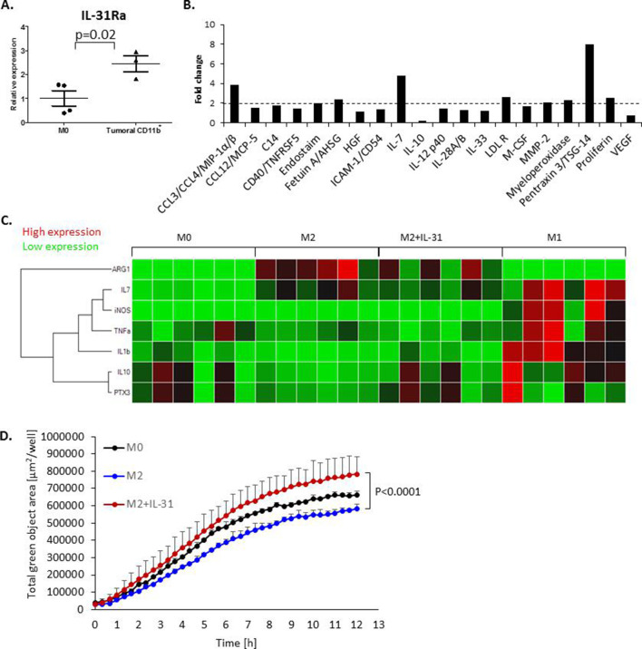
Figure 4.
IL-31 induces a proinflammatory state in macrophages and enhances macrophage phagocytic activity. (A) The level of IL-31Ra mRNA was measured by RT-qPCR in naïve M0 BMDMs and in CD11b+ myeloid cells isolated from PyMT tumors (n=3–4 biological repeats). (B, C) BMDMs were isolated from naïve female C57BL/6 mice and skewed toward the M2 phenotype (by IL-4 treatment), the M1 phenotype (by interferon-γ treatment) or left as the M0 phenotype (no additives). M2 BMDMs were treated with 100 ng/mL IL-31 or left untreated (control). The levels of various secreted proteins in the conditioned medium were measured. Data are presented as fold change relative to control. Dashed line represents all proteins which displayed over twofold increase (B). The mRNA expression levels of various inflammatory and anti-inflammatory factors were evaluated in untreated M0, M1 and M2 BMDMs, and IL-31-treated M2 BMDMs (M2+IL-31). Shown is a heat map of normalized values (C). (D) The phagocytic activity of untreated M0, untreated M2 and IL-31-treated M2 macrophages (M2+IL-31) was measured by IncuCyte. Statistical significance was assessed by unpaired two-tailed t-test. Significant p values are shown. BMDMs, bone marrow-derived macrophages; IL, interleukin; IL-31Ra, IL-31 receptor A; RT-qPCR, real-time quantitative PCR; AHSG, alpha 2-heremans schmid glycoprotein; ICAM-1, intercellular adhesion molecule 1; HGF. hepatopoietin-A; LDL R, low density lipoprotein receptor; M-CSF, macrophage colony-stimulating factor; MMP-2, matrix metalloproteinase-2; TSG-14, tumor necrosis factor-inducible gene 14; VEGF, vascular endothelial growth factor; iNOS, inducible nitric oxide synthase; TNF-a, tumor necrosis factor alpha; PTX3, pentraxin 3.

