Figure 4.
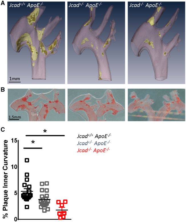
Loss of Jcad causes a significant reduction in atherosclerosis in the aortic arch inner curvature. (A) Representative three-dimensional renders generated from microCT images of atherosclerosis plaques in the aortic arch of hyperlipidaemic female wild-type (Jcad+/+ ApoE−/−), heterozygous (Jcad+/− ApoE−/−), and homozygous (Jcad−/−ApoE−/−) knock out mice fed a high-fat diet for 10 weeks. Vessel wall is coloured pink, plaque yellow, and necrotic core dark yellow, scale bar = 1mm. (B) Representative images of the aortic arch stained for atherosclerotic plaques using Oil Red O lipid staining (plaque stains red; scale bar = 1.5 mm). (C) Quantification of en face plaque area in the inner curvature of the aortic arch reveals a significant reduction in plaque area in the inner curvature of Jcad+/−ApoE−/− and Jcad−/−ApoE−/− versus Jcad+/+ ApoE−/− mice (*P < 0.05, Kruskal–Wallis test, n = 18–7 per group). Data are expressed as the mean ± SEM, each point represents an individual animal.

