
Fig. 1.
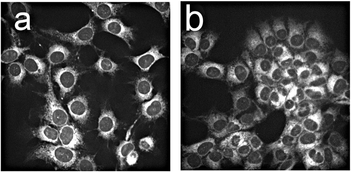
Fluorescence localization in OVCAR5 cells of BPD dissolved in DMF (a) or delivered as the lipo-BPD formulation termed BPDf (b). The extracellular concentration of BPD was 0.5 μM with images acquired after 1 hour incubations.
Official websites use .gov
A
.gov website belongs to an official
government organization in the United States.
Secure .gov websites use HTTPS
A lock (
) or https:// means you've safely
connected to the .gov website. Share sensitive
information only on official, secure websites.
Fluorescence localization in OVCAR5 cells of BPD dissolved in DMF (a) or delivered as the lipo-BPD formulation termed BPDf (b). The extracellular concentration of BPD was 0.5 μM with images acquired after 1 hour incubations.