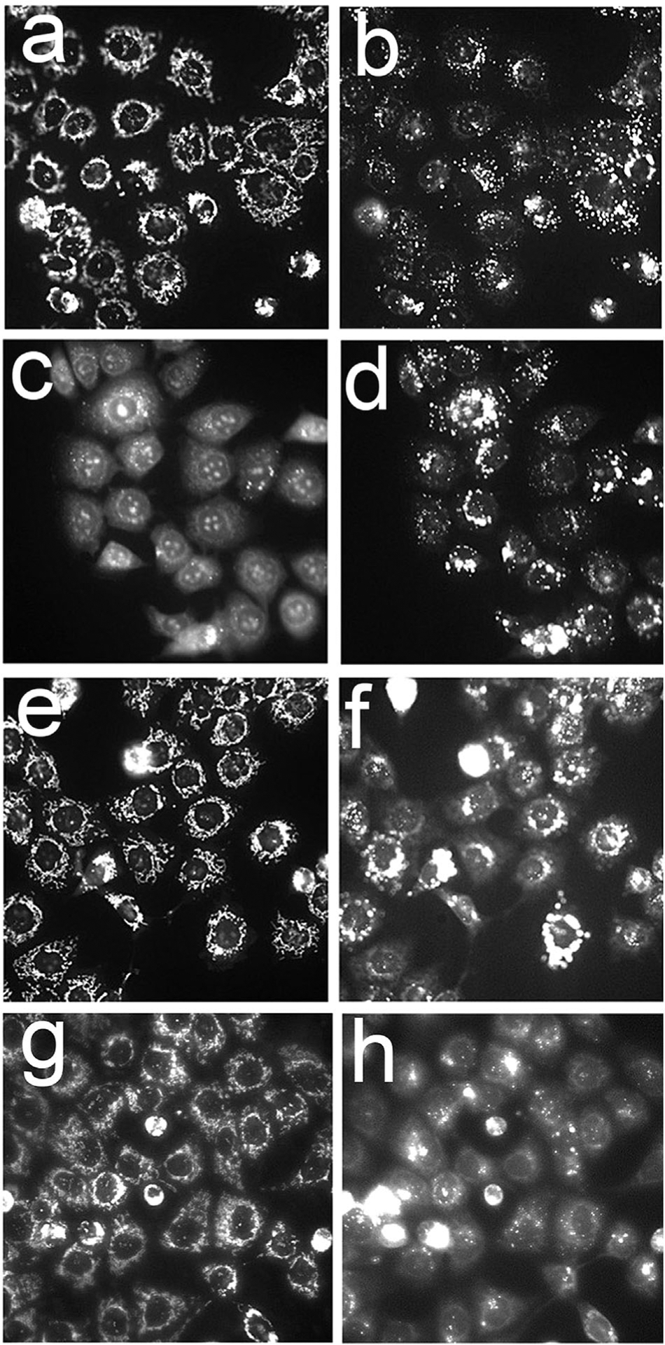
Fig. 3.

Effects of different BPD formulations on the mitochondrial membrane potential probed with MTO (a,c,e,g) and the lysosomal pH gradient as detected by LTG (b,d,f,h) directly after irradiation (690 ± 10 nm, 600 mJ/sq cm). a,b = untreated cells; c,d = BPDf; e,f = BPDa+c; g,h = BPDa−c The extracellular BPD concentration was 0.05 μM (BPDf) or 0.5 μM (BPDa). Cells were incubated with the liposomal formulations for 3 hours before irradiation.
